

Table of Contents
- Summary & Introduction
- Epifaunal Worm Tubes on Lower Lias Ammonites – Results
- Epifaunal Worm Tubes on Lower Lias Ammonites – Detailed Observations and interpretation
- Epifaunal Worm Tubes on Lower Lias Ammonites – Discussion
- Epifaunal Worm Tubes on Lower Lias Ammonites – Conclusions and References
3. Results
Over 40 ammonites with worm tubes attached have been considered in detail (Table 1), of which 38 are pyritized Promicroceras (e.g., Figs 2-7). In addition, seven Promicroceras with fragments of worm tubes attached were collected that are too poorly preserved for analysis.
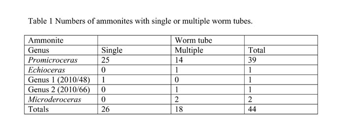
It is significant that, for the ammonites with single (Figs 2-4) or multiple (Figs 5-7) peripheral tubes, not one worm grew counter to the direction of growth of the ammonite to which it was attached. In summary, the most common association among our specimens is of a single, peripheral worm tube (Figs 2-4) growing on the ammonite Promicroceras. In many cases the ammonite eventually grew over the worm tube, clearly indicating that the ammonite was alive while the worm tube was growing. We regard this most common association as the ‘standard pattern’ and discuss its significance below (Section 4.1).
-
- Fig. 2. Left side of LYMPH 2010/29 a Promicroceras with single, peripheral worm tube. The worm tube starts just above (o) in the umbilical seam on the left side of the ammonite, follows the umbilical seam for over half a whorl in the same direction as the ammonite growth and then crosses the penultimate whorl where it is overgrown by the ammonite. It continues around the periphery of the ammonite and terminates well short of the ammonite aperture, but is incomplete. Scale bar = 5 mm.

-

- Fig. 3. Left side of LYMPH 2010/48 a Promicroceras with a worm tube emerging from beneath the ammonite and growing around much of the periphery of the ammonite. Scale bar = 5 mm.

-

- Fig. 4. Left side of LYMPH 2010/32 to show worm tube emerging from beneath the ammonite and continuing along the venter for almost a whorl. Scale bar = 5 mm.

-
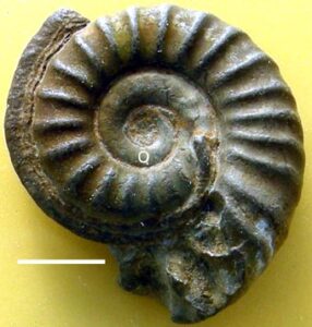
- Fig. 5. A, left and B, right sides of LYMPH 2010/53 a Promicroceras with four peripheral worm tubes (1-4). Scale bar = 5 mm.

-
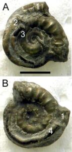
- Fig. 6. A, left and B, right sides of LYMPH 2010/38, a small Promicroceras with three peripheral worm tubes. The secondary worm tubes nestle in the groove between the primary worm tube and the ammonite shell. Scale bar = 5 mm.

-

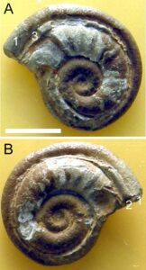
- Fig. 7. Right side of LYMPH 2010/50 a Promicroceras with two peripheral worm tubes, one of which attached on the right side. Scale bar = 5 mm.
